What is Venous Ultrasound?
Venous ultrasound, also known as a Doppler vascular ultrasound, is an ultrasound exam that looks at the vascular system. The vascular or circulatory system consists of the veins and arteries that carry blood throughout the body. This type of ultrasound uses Doppler technology to create color images and videos of what is happening inside the body. This is accomplished with harmless, high-frequency soundwaves that show blood flow speed and direction within the veins.
Venous ultrasound exams are used for both diagnostic and monitoring purposes. These may include vein mapping, identifying blood clots, and post-surgery monitoring.
Venous Ultrasound for Diagnosis and Monitoring
Venous ultrasound can be a critical diagnostic tool for various conditions such as blood clots and vascular disease. Examples of vascular disease include the following:
- Atherosclerosis (arterial blockages)
- A buildup of fatty deposits within the arteries.
- Abdominal Aortic Aneurysm (AAA)
- A bulge in the aorta (the largest artery in the body) within the abdomen.
- Carotid Artery Disease
- A buildup of plaque within the carotid arteries is located on either side of the neck.
- Deep Vein Thrombosis (blood clots)
- A blood clot within the veins. Usually present in the legs.
- Varicose Veins
- Swollen or enlarged veins are caused by weak valves. Usually present in the legs.
Venous ultrasound is also used for vein mapping. The vein mapping process uses venous ultrasound to take pictures of the veins and arteries to help create a ‘map’ of the blood vessels within the body. Vein mapping is used to help guide medical procedures, including:
- Bypass Surgery: For bypass surgery involving the blood vessels, the procedure is to take a portion of a healthy blood vessel from one part of the body and use it in a different part of the body to ‘bypass’ a blocked artery and reroute blood flow. Two main types of venous bypass surgery include coronary artery bypass and peripheral vascular bypass
- Kidney Dialysis: Dialysis is a treatment for kidney failure. When functioning properly, the kidneys filter toxins and waste from the blood. During dialysis, blood is removed from the body and filtered using a machine. In some cases, this process is assisted by connecting an artery and vein in the arm to help remove the blood and deliver it back to the body. Vein mapping determines which veins and arteries to use in the procedure.
Venous ultrasound can also be used as a monitoring tool in a post-diagnostic or post-surgery environment. For example, suppose surgery is not required in the case of an abdominal aortic aneurysm (AAA). In that case, an ultrasound exam may monitor the aneurysm over time and ensure it doesn’t get larger. Examples of post-surgery ultrasound exams include monitoring the status of a stent implanted to treat an arterial blockage or to ensure an arterial graft from a bypass procedure is not rejected.
Venous Ultrasound Exam Process
A doctor will refer the patient for an exam if a venous ultrasound is deemed necessary for diagnostic, surgical, or monitoring purposes. Exams are performed by Registered Vascular Technologists (RVTs) who have been trained and certified in vascular ultrasound. Here is an example of a typical examination:
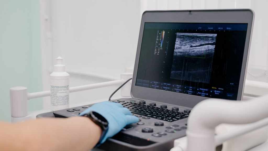
- The ultrasound machine consists of a video monitor, console, and a handheld device called a transducer, which emits high-frequency soundwaves that help create images of the veins and arteries.
- The patient will be asked to lie on a table for an exam. Depending on the body area that needs to be examined, the patient may be asked to change into a medical gown onsite.
- The RVT will apply a small amount of gel to the ultrasound transducer, which helps soundwaves travel better by eliminating air pockets. The RVT will then use the transducer to apply pressure to the skin of the examined area. This gentle compression can help determine if there are abnormalities in the veins (e.g., veins that cannot be easily compressed may indicate a blockage).
- Physicians who are trained to read and interpret ultrasound images will then review the results and send them to the patient’s physician for final diagnosis or analysis. At this point, the patient’s physician can make informed treatment or surgical recommendations as necessary.
Overall, venous ultrasound is an accurate and non-invasive way to diagnose various conditions, provide vein mapping for surgical procedures, and may also be used to monitor the vascular system after diagnosis or surgery. Venous ultrasound exams are painless and pose no known health risks. For these reasons, it is useful in various inpatient and outpatient settings.
Guest Contributor: Jordan Galerkin
Sources:
- Arterial & Venous Mapping. Cleveland Clinic. https://my.clevelandclinic.org/health/diagnostics/17607-arterial–venous-mapping
- Dialysis. Cleveland Clinic. https://my.clevelandclinic.org/health/treatments/14618-dialysis
- Ultrasound. Mayo Clinic. https://www.mayoclinic.org/tests-procedures/ultrasound/about/pac-20395177
- What to Know About Vascular Disease. Medical News Today. https://www.medicalnewstoday.com/articles/vascular-disease
