A carotid duplex ultrasound is a type of Doppler ultrasound. The carotid duplex ultrasound is an exam used to help diagnose carotid artery disease. Carotid duplex ultrasound is just one type of duplex ultrasound. Other types of duplex ultrasounds image other areas of the body, such as the kidneys (renal duplex ultrasound).
Doppler ultrasound technology can create color images and videos of what is going on inside the body using high-frequency soundwaves. These soundwaves can show both direction and speed of blood flow within the veins and arteries. Duplex ultrasounds also show images of tissues surrounding the veins and arteries.
Doppler ultrasound exams can be a critical diagnostic tool. Since they can determine speed and direction of blood flow, they can detect potential blockages in the arteries, which can be fatal. These blockages may be caused by blood clots or plaque buildup. The sooner these issues can be identified, the sooner treatment can take place.
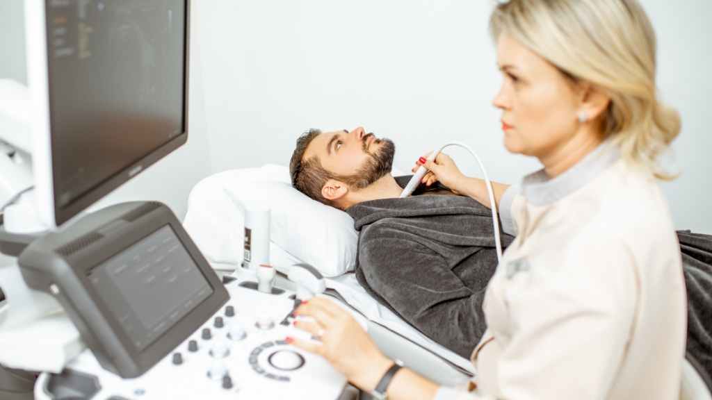
The process of undergoing a doppler or duplex ultrasound exam is a simple one. The ultrasound machine is made up of three basic parts; a video monitor, a console, and a handheld device called a transducer, which is the part that emits the high-frequency soundwaves. It’s important to understand that ultrasound exams are painless, pose no known health risks, and can be a life-saving diagnostic tool.
Sources:
- Types of Ultrasound. Ultrasound Quotes. https://www.ultrasoundquotes.com/blog/doppler-vs-duplex/
- Ultrasound. Mayo Clinic. https://www.mayoclinic.org/tests-procedures/ultrasound/about/pac-20395177
