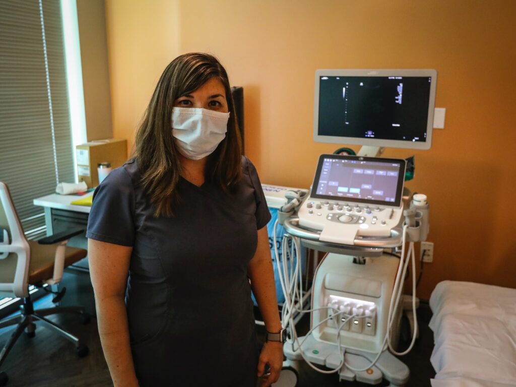
You may have had a vascular ultrasound if you’ve ever wondered why your foot or ankle swells up and bruises easily or have pain in your leg below the knee that doesn’t seem to go away with rest or ice. Vascular ultrasounds are one of the most common scans by radiologists across the country. They can help detect blood clots, inflammation, atherosclerosis, and many other conditions that can affect the blood vessels of your leg.

What is a Vascular Ultrasound
A vascular ultrasound is a diagnostic test that uses high-frequency sound waves to produce images of the blood vessels. The test can be used to detect a variety of conditions, including blockages, aneurysms, and other abnormalities. The test is noninvasive and does not require any special preparation. A small device, called a transducer, is placed on the skin over the area of interest. The transducer emits sound waves that bounce off the blood vessels and are then converted into images. The images are displayed on a monitor for the doctor to review.
Who Needs A Doppler Exam
A Doppler exam may be recommended if you have symptoms of poor circulation, such as pain in your legs when walking. Your doctor may also recommend an abdominal aortic aneurysm screening if you’re at risk for this condition. Other reasons you may need a Doppler exam include:
- You have high blood pressure
- You have diabetes
- You’ve had a heart attack or stroke
- You have hardening of the arteries (atherosclerosis)
- You have a family history of aneurysms
Why A Doppler Exam Is Used
A Doppler exam can be used to measure how fast blood is moving through your arteries. This information can help your doctor detect an abdominal aortic aneurysm (AAA). An AAA is a bulge in the aorta, the large blood vessel that carries blood from your heart to the rest of your body. A small AAA may not cause any symptoms. But a large AAA can rupture, causing life-threatening bleeding. A Doppler exam can also screen for other conditions, such as the hardening of the arteries (atherosclerosis) or blockages.
How To Prepare For A Doppler Exam
Before your exam, you will likely need to fast for four to six hours. This means no eating or drinking during that time. You may also be asked to drink plenty of water before the exam, so your bladder is full. This is because a full bladder pushes the intestines out of the way, making it easier to see your abdominal aorta. You will likely be asked to wear a gown during the exam. The ultrasound technician will then apply gel to your stomach area and place the transducer (the device used to send sound waves into your body) on top of it. He or she will use this device to send sound waves into different parts of your abdomen to create images that can show any abnormalities. The technician may also use devices such as a Doppler probe to check how well blood flows through the arteries near the kidneys, spleen, liver, pancreas, heart, and brain.
Things to Know After Your Examination
After your examination, the sonographer will give you a brief description of what was found. A radiologist will read the images and send a report to your referring physician. Your physician will go over the findings with you and explain what, if any, treatment is necessary. abdominal aortic aneurysm screening is just one of many uses for vascular ultrasound. The test can also evaluate blood flow, look for artery blockages, or assess damage after a heart attack.
Sources:
- Ultrasound – Vascular. Radiological Society of North America, Inc. https://www.radiologyinfo.org/en/info/vascularus
- Frequently Asked Questions or What Can You Expect? UC Davis Health. https://health.ucdavis.edu/radiology/mymri/myexam/myvl/FAQ/vasfaq.html
- Vascular Ultrasound. Cleveland Clinic. https://my.clevelandclinic.org/health/diagnostics/17606-vascular-ultrasound
